Understanding the immune system
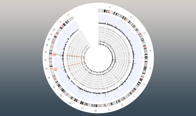
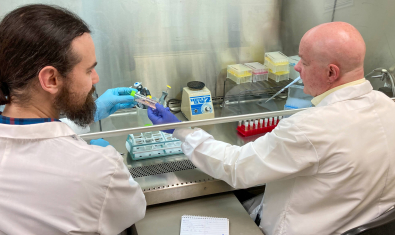
Scientists in our Center for Fundamental Immunology conduct research to better understand how the immune system works in health and disease. We use advanced tools, technologies and methods to study the genes, cells and molecules in the immune system, often in partnership with BRI’s Center for Systems Immunology. Our work includes building complex models of autoimmune and inflammatory diseases to study them in precise detail. We also examine samples from people with and without immune system diseases to gain insight into what goes wrong in disease and to identify possible targets for new therapies.
We work closely with BRI’s Centers for Translational Immunology and Interventional Immunology. These groups build on our fundamental insights, seeking out applications for our research in human immunology and patient care. We also collaborate with these centers to analyze clinical trial data to better understand the mechanistic basis for new immune therapies.
Daniel J. Campbell, PhD
Labs in the Center for Fundamental Immunology

Bettelli Lab
The main focus of the Bettelli Lab is to identify the cell types of the immune system and mechanisms, which induce and regulate the development of autoimmunity.
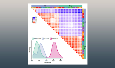
Campbell Lab
The Campbell laboratory is interested in understanding the basis for T cell activation, function and tolerance.
Hamerman Lab
The lab is interested in understanding how myeloid cells contribute to both productive and pathological immune responses during infection, inflammation, and autoimmune diseases.
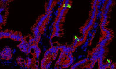
Harrison Lab
The Harrison Lab studies the mechanisms controlling host-microbe interactions at barrier tissues, primarily the skin and the gut with the goal to understand how these immune cells promote barrier tissue integrity and repair, and to understand how this goes awry during disease.
Lacy-Hulbert Lab
Researchers in the Lacy-Hulbert Lab study how components of the immune system work together to defend against harmful invaders while avoiding damage to healthy tissues or harmless microbes.
Morawski Lab
The Morawski lab studies the adaptive immune response occurring during human inflammatory and autoimmune diseases of the skin and other barrier tissues.
Ziegler Lab
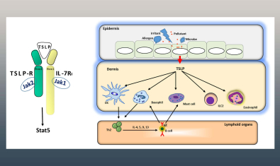
The Ziegler laboratory is investigating the role of the epithelial cytokines TSLP and IL-33 in regulating protective responses at mucosal barrier surfaces such as the respiratory and gastrointestinal tracts.
Gut Immunity Program
The Gut Immunity Program studies how immune responses in the gut go wrong and lead to inflammatory bowel diseases (IBD) such as, celiac disease, Crohn's & colitis and other autoimmune conditions.
Researchers use model systems, biorepository samples and clinical studies to understand how immune responses go awry. They also study the gut microbiome and how immune responses in the gut contribute to other autoimmune diseases.
News
Regulatory T Cell-Boosting Therapy Impacts Flu Responses — For Better or Worse — Depending on Timing
Read More
New Research Reveals Rheumatoid Arthritis Begins Years Before Symptoms
Read More ➡