Blog Main Scientific Organoid Blue Green
September 21, 2023

Revolutionary Microscope to Fuel Immunology Breakthroughs

What if we could take a blood sample from a person with rheumatoid arthritis, zero in on the immune cells that cause the disease, and watch what they’re doing in real-time video? And what if we could could separately test over 300 different treatments on those cells, aiming to slow down their attack and gain crucial insight into which therapy works best and why?

Thanks to a state-of-the-art new microscope, BRI scientists can now do that and so much more.

Caroline  Stefani
Caroline Stefani, PhD, Principal Investigator and Imaging Core Manager

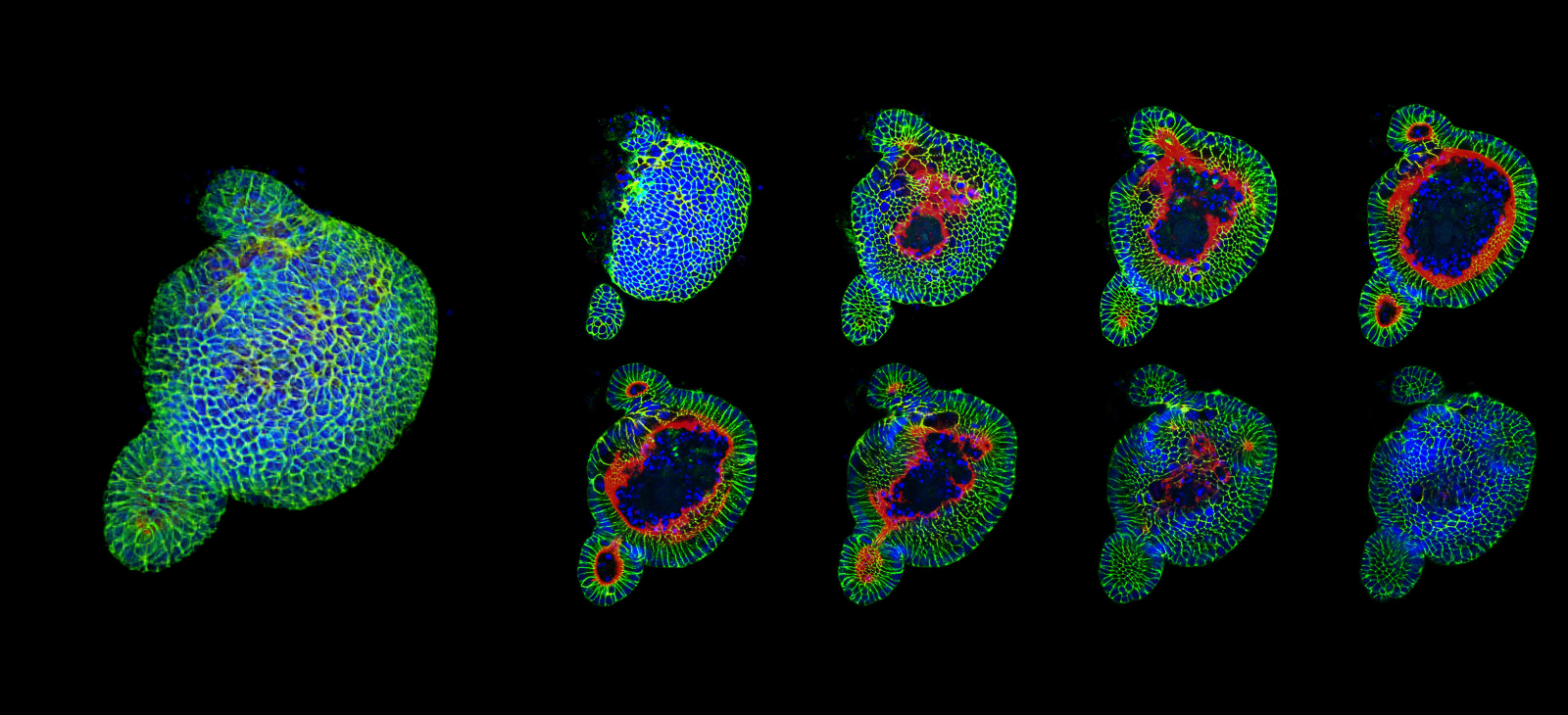
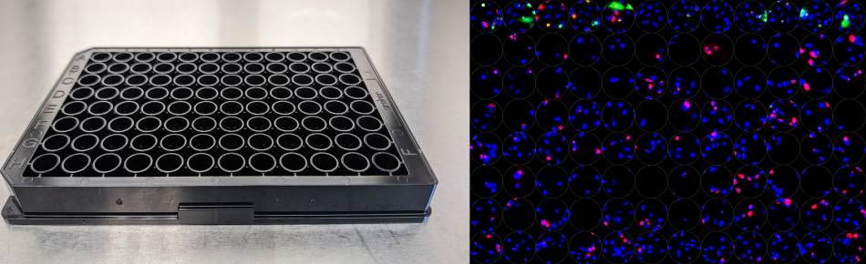
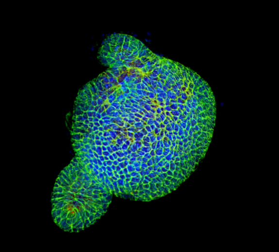
“This is now the most powerful microscope we have at BRI, and BRI is one of only three academic institutions in the Pacific Northwest with this technology,” says Caroline Stefani, PhD, a systems immunologist who leads BRI’s Imaging Core. “These high-end technologies enable us to go faster, answer bigger questions, and branch out into new and exciting areas of research.”

Dr. Stefani is an expert in using advanced imaging tools to extract as much data from blood and tissue samples as possible. With this new tool, she’ll partner with scientists across BRI to dig deeper into existing research questions and take on new challenges in immune disease research. The following are highlights of how it will help advance research.

Examining Eight Markers (Data Points) at the Same Time

The new microscope increases our ability to study as many as eight markers from one sample; our previous tool could only gather four. With this added capability, we can gather significantly more information from the sample of blood or tissue we’re studying.

Full-Width Scientific Sample Sections

Viewing Every Section of a Sample

Looking at flat images can help scientists make good insights. But examining cross-sectional images is even better. Think of it this way: The machine could take a picture of a tomato and then create a detailed picture of individual tomato slices. This allows scientists to gather more detail and look at samples from different angles. This is particularly consequential when we use it on organoids — lifelike models of human guts or lungs — and study each layer of tissue in precise detail.

Imaging up to 384 Wells

Graphic 96-Well Plateview

Our new microscope can image up to 384 “wells” at the same time. Individual wells enable scientists to see how cells behave under different circumstances — for example, how cells respond to different medicines or different doses of medicine. This new tool can also record videos of cells over minutes, hours and even days. With the ability to collect more data and more details, we can accelerate the pace of our understanding of diseases, the immune system and health.

Peter Morawski
Peter Morawski, PhD, Principal Investigator

Making Dreams Reality

Skin immunology researcher Peter Morawski, PhD, can hardly wait to use this microscope. He studies how skin immune cells protect against infections and help heal wounds — and why these cells sometimes go into overdrive and lead to diseases like psoriasis and scleroderma.

In the past, this work would have required using several different tools to run several different experiments and then integrating the results at the end. The new microscope will let him run all of the experiments in one place and collect significantly more data about the skin and tissue biology. The ability to take videos will allow him to see how the cells interact and respond over time.

“We’ll examine skin immunology in a whole new way,” Dr. Morawski says. “It’s unexplored territory, and it’s really exciting because we don’t know what we’ll find.”

Dr. Morawski’s ultimate goal would be to run a complex study examining how immune cells respond when skin cells die. Sometimes skin cells die in a very controlled process that makes space for new cells. And sometimes it’s a more inflammatory and potentially problematic response after an infection.

Scientists don’t fully understand how each kind of cell death triggers different immune responses — and if something about these processes could explain why immune cells go into overdrive, leading to diseases like scleroderma and lupus.

“This advanced technology can provide crucial insight into this question and many more,” Dr. Morawski says. “It helps turn pipe dreams into reality. It enables us to answer the most pressing biological questions, and move closer to discoveries that can help people.”

Daniel Campbell
Dan Campbell, PhD, Director of BRI's Center for Translational Immunology

Philanthropy Makes a Big Impact

Technologies like this microscope are vital to BRI’s research, but they are expensive. Donors fill a crucial gap in acquiring state-of-the-art tools because research grants don’t typically cover them. BRI would not have been able to purchase this tool without a generous grant from the M.J. Murdock Charitable Trust.

“Investing in tools and technology opens the door to previously unexplored ways of looking at disease. It’s one of the best ways to have a broad, long-standing impact on our research,” says Dan Campbell, PhD, director of BRI’s Center for Fundamental Immunology. “The Murdock Trust has been fueling cutting-edge research at BRI for 20 years and we’re immensely grateful. Their support enables us to look deeper into the immune system and identify new ways to intervene in disease.”

Donate Now

Immuno-what? Hear the latest from BRI

Keep up to date on our latest research, new clinical trials and exciting publications.