Microscopy & Imaging
The Imaging core is composed of 6 instruments and 2 workstations. In addition to hands-on training and assisted-use options for all our machines, we offer consultation services to both internal and external research groups.
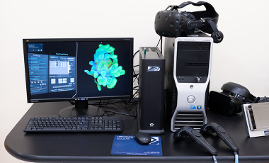
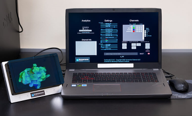
We provide consulting on experiment design and troubleshooting. We are also available to perform a large panel of image analysis: high-content, morphological analysis, chemotaxis and other live imaging, 3D and Virtual Reality, screening, etc.
Available Imaging Equipment
We have the following equipment available for use.
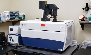
ImageXpress Confocal HT.ai High-Content Imaging System
The ImageXpress confocal HT .ai, is an automated imaging system containing a spinning disk confocal with seven laser light sources and eight imaging channels enabling highly multiplexed assays (e.g. screening, CellPainting). Its fully controlled environment allows for live imaging of cells or tissue in brightfield, phase contrast or fluorescence. Its equipped with a TL tower and multi-slide holder combined with the automated stitching function for slide scanning of H and E or fluorescent staining.
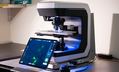
Echo Revolve upright and inverted Discover
This microscope can be used to image specimens mounted on slides or unmounted on dishes. The microscope is connected to an iPad, making it easy to use and collaborate. The entire microscope rotates from inverted to upright.
Lenses: The microscope is equipped with 4X-10X-20X-40X and 60X. All of these lenses are "dry", i.e., they are not to be used with immersion oil.
Illumination
- Transillumination in brightfield, and phase-contrast
- Fluorescence epi-illumination for blue (e.g., DAPI), green (e.g., FITC/GFP), red (e.g., TRITC/Texas Red) and far red (e.g Cy5) fluorescent dyes.
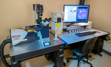
SP5 Leica confocal
This inverted microscope can be used to image specimens mounted on slides or in coverslip-bottomed chamber slides or dishes. Magnification range is 100X–630X as viewed through the eyepieces. Higher magnifications can be achieved in confocal mode by use of digital enlargement. The microscope is fully automated and can be controlled by sets of buttons on the microscope itself or by the confocal scan software. The Live Cell™ imaging system (shown at right) allows investigators to culture living cells or tissue explants on the microscope stage under controlled temperature, CO2, and humidity for at least 48 h. This system can be used for image acquisition in real-time and in time-lapse.
Lenses: The microscope is equipped with 10X dry, 20X dry, 20X multi-immersion, 40X oil immersion, and 63X glycerol immersion objective lenses. The 20X multi-immersion lens can be used dry, but works best with water. It has a correction collar that can be set for air (O), water (W), oil (OIL), and glycerol (GLYC).
Illumination
- A standard transmitted light lamp for viewing specimens through the microscope eyepieces via brightfield or differential interference contrast (DIC) (Nomarski) modes.
- A fluorescent lamp (Leica EL 6000) for viewing specimens through the microscope eyepieces via 3-color (red, green, blue) epi-fluorescence.
- Four lasers (blue, green, orange, red) provide epifluorescence and DIC illumination in scanning confocal mode. A total of 8 laser lines (458, 476, 488, 496, 514, 543, 594, and 633 nm) are available for dye excitation.
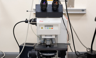
DMR upright Leica
This microscope can be used to image specimens mounted on slides. Magnification range is 25X–1,008X as viewed through the eyepieces.
Lenses: 2.5X, 10X, 20X, 40X (two lenses), and 63X short working distance objective lenses. All of these lenses are "dry", i.e., they are not to be used with immersion oil.
Illumination
- Transillumination in brightfield, darkfield, phase-contrast, polarized light, and Nomarski modes.
- Fluorescence epi-illumination for blue (e.g., DAPI), green (e.g., FITC/GFP), and red (e.g., TRITC/Texas Red) fluorescent dyes.
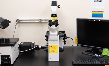
DMIRB inverted Leica
This microscope can be used to image cell preparations in tissue culture plates, dishes, or flasks. You can also image specimens mounted on slides if the slides are turned upside down. Magnification range is 25X–945X as viewed through the eyepieces.
Lenses: 2.5X, 10X, 20X, 40X, and 63X long working distance objective lenses. The 20X, 40X, and 63X lenses have correction collars to optimize image sharpness. None of the lenses are oil immersion.
Illumination
- Transillumination in brightfield, darkfield, phase-contrast, polarized light, and Nomarski modes.
- Fluorescence epi-illumination for blue (e.g., DAPI), green (e.g., FITC/GFP), and red (e.g., TRITC/Texas Red) fluorescent dyes.
- The fluorescence illuminator uses a liquid light guide that does not need alignment. The intensity of the fluorescence lamp can be varied and its light can be cut off to the microscope with a shutter button on the illuminator housing.
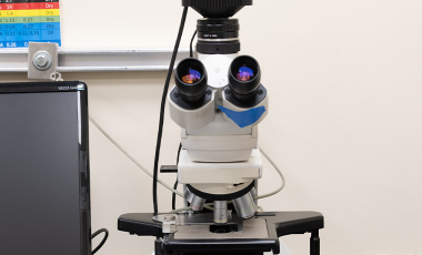
MZFL III Stereo
This microscope provides low to midrange magnification (5X–160X as viewed through the eyepieces) with a long depth of field. It is well suited for macro-scale imaging of whole or dissected organs or tumors from small animals, whole-mount tissue culture preparations, cultures transfected with fluorescent reporters, and small biomedical constructs or devices.
Lenses
- 0.63X long working distance objective lens.
- 1.00X intermediate working distance objective lens.
- 1.60X short working distance objective lens.
- 0.8X–10X zoom intermediate lenses.
Illumination
- Brightfield, darkfield, and semi-darkfield transillumination modes.
- White light epi-illumination by top (fiber optic) or dual sidelights. Two types of dual sidelight are available: (1) conventional filament lamps and (2) high-intensity LED lamps.
- Fluorescence epi-illumination for blue (e.g., DAPI), green (e.g., FITC/GFP), and red (e.g., TRITC/Texas Red) fluorescent dyes.
DM 2500 3-Head
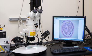
This microscope can be used to image specimens mounted on slides. The instrument has one set of binocular eyepieces and three LCD monitors. The monitors receive identical feeds from the computer and can be used to view live or captured images from the digital camera. The magnification range of the microscope is 12.5X–630X as viewed through the eyepieces. This microscope is the first choice for imaging specimens stained with histological dyes (e.g., hematoxylin & eosin, Masson's, Movat's, PAS, etc.).
Lenses: .25X, 5X, 10X, 20X, 40X, and 63X short working distance objective lenses These lenses are not to be used with oil.
Illumination: Transillumination in brightfield, darkfield, phase-contrast, and polarized light modes.