Introduction
Histology Core at BRI
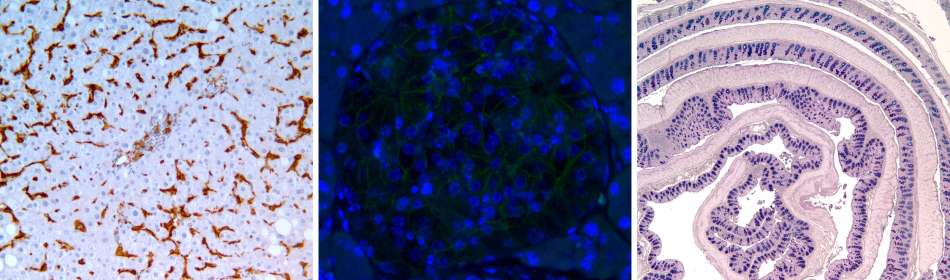
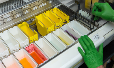
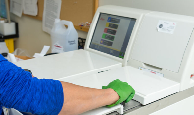
The Histology Core Laboratory provides comprehensive histological services to both BRI scientists and the surrounding biotech community, from external academic investigators to large multinational biotech companies.
Managed by Dr. Pamela Johnson, the core facility utilizes high-throughput automated equipment for tissue infiltration, embedment in paraffin or OCT, sectioning (microtomy, cryotomy), dye-based and immunohistochemical staining, spatial transcriptomics, tissue matrix array, and cover-slipping. Leveraging Dr. Johnson’s 25+ years’ experience, the facility also specializes in troubleshooting and protocol development.
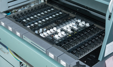
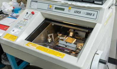
Histology Core Equipment
Histology Core Lab Members

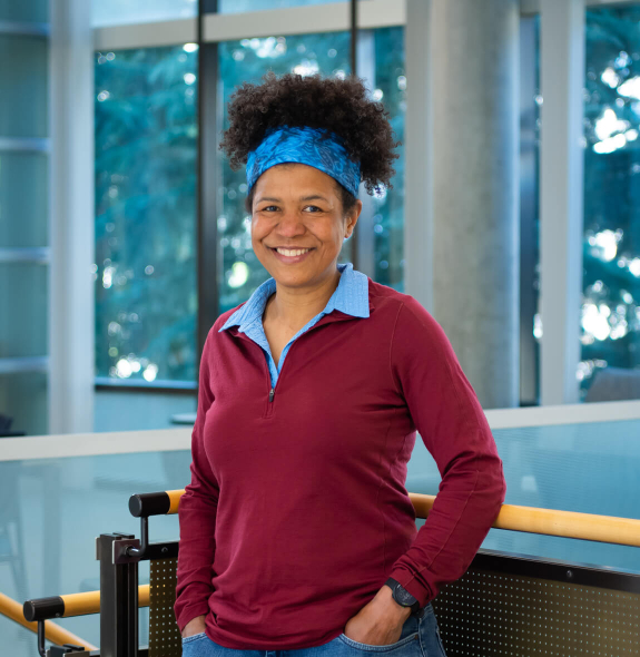
Riley Robles
Research Technician, Histology Core Lab

