Blog Main Image - Scientific Proteoglycans Excessive Deposition Vascular Disease
August 26, 2019

Using Virtual Reality to See Inside Cells

In 2016, two of BRI's information technology experts Garrett Wright and Tom Skillman had a conversation that sparked a novel question: Could virtual reality (VR) headsets let scientists step inside cells and view them in greater detail than ever before?

Featured BRI Tom Skillman Caroline Stefani

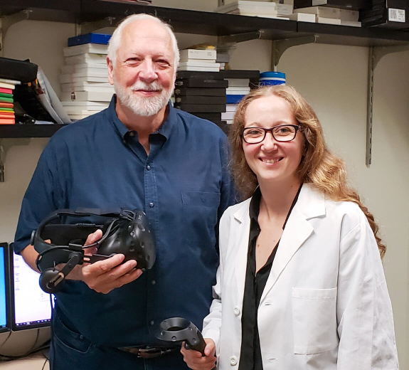
When Skillman shared this idea with Caroline Stefani, PhD, a senior postdoctoral associate in the Lacy-Hulbert & Stuart Laboratories, she was intrigued — and her expertise in 3D microscopes made her the perfect research partner to help create this VR technology.

Fast-forward three years, and Dr. Stefani and Skillman (BRI’s former director of research technology who now leads his own company, Immersive Science) are making the technology even more powerful — through a grant from the Bill & Melinda Gates Foundation and a partnership with Yongxin (Leon) Zhao, PhD, at Carnegie Mellon University (CMU). 

“This VR tool will give us new insights into autoimmune diseases, infections and more,” Dr. Stefani says, “and we’re working to make it accessible for scientists everywhere.” 

Better Than Microscopes

Looking at cells using traditional microscopy is like looking at a 2D picture of a city while knowing the actual city is 3D. The new VR software, called ConfocalVR, allows scientists to visually step inside tissues and cells and see things they never saw in 2D.

“We have scientists who’ve been studying the same types of proteins for 40 years, and they’re blown away when they see them in VR,” Dr. Lacy-Hulbert says.

Gates Foundation Grant

The grant and partnership arose from Skillman and Dr. Stefani presenting their software at the 2018 Grand Challenges Annual Meeting (GCAM) to the Gates Foundation and other members of the global Grand Challenges community. Scientist GCAM attendees were invited to view their own data in VR. The technology intrigued CMU’s Dr. Zhao — who has been pioneering his own brainchild, called expansion microbiology.

Featured Bio Yongxin Zhou

Right now, light microscopes can only zoom in so far before an image gets blurry and grainy. This means it’s hard to study especially tiny features of cells, particularly features of microbial cells — those that come from bacteria and other invaders. To overcome this, Dr. Zhao started using a special gel to expand microbial cells to 100 times their actual size (picture an ant becoming as big as a rhinoceros).

“This is the future of complex imaging,” Dr. Zhao says. “It’s like watching a movie in IMAX versus watching it on your phone.” 

Global Impact

BRI is already sending cells from our biorepositories to CMU. Dr. Zhao’s team enlarges them, tags key proteins with different colors, and scans them using a special 3D microscope. Then CMU’s team sends the digital image data back to BRI for analysis.

Our team will use software being specifically developed for this project to accelerate our understanding of this data using VR. 

“In tuberculosis, for example, microbes go into your cells,” Dr. Stefani says. “Now, we can see exactly where they go and what they do.” 

The Gates grant requires the researchers to make this expanded technology, called ExMicroVR, affordable and accessible. Dr. Stefani envisions a future where scientists around the world can download it.

Researchers on the frontlines of infectious disease research in Africa could then upload their data and examine it in real time with other experts around the world. 

“This tool could help scientists everywhere,” Dr. Stefani says. “And it all started because BRI believes in collaboration and encourages scientists to pursue outside-the-box ideas.”

Immuno-what? Hear the latest from BRI

Keep up to date on our latest research, new clinical trials and exciting publications.