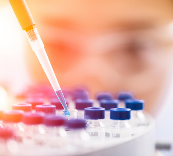
Blog Main Image - Researcher Woman Pipetting Samples Blue
April 1, 2024

New Frontiers in Immunology: Studying Diseases in the Tissues Where They Happen

Traditionally, scientists have studied immune system diseases by looking at blood samples. But they’ve also known studying the actual tissue that those diseases affect could provide even more information. The problem is, tissue is hard to come by — it’s more feasible to collect and study blood samples than it is to do the same with tissue from joints or the pancreas. 

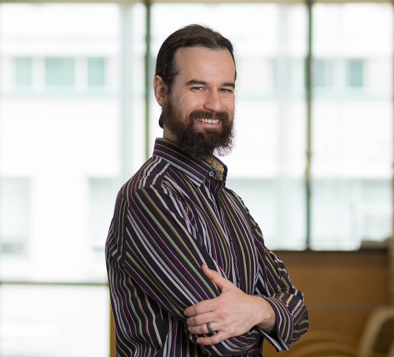
Eddie James
Eddie James, Principal Investigator, James Lab

“Blood samples are informative but they don’t tell the whole story,” says Eddie James, PhD. “The real action is in the tissue, but we haven’t really had access to it in the past.”

Dr. James is one of many BRI scientists working to change that by finding new ways to study tissue — from studying donated tissue from people who’ve had joint replacements to creating engineered models of human skin. This work is placing BRI at the forefront of a field called tissue immunology. And it’s leading to insights that could transform our ability to predict, prevent, reverse and cure immune system disease.

“We’re learning that immune cells behave differently based on the environment of the tissues they end up in. Think of it like cars that are made for different environments — some are designed to go fast on a racetrack, while others are for off-roading,” says Peter Morawski, PhD. “Studying how cells adapt to their environment provides a lot of insight into what’s happening in disease, and we’re just starting to scratch the surface.” 

Getting to the Root of RA

Dr. James is on a quest to understand exactly how and why rheumatoid arthritis (RA) happens. To do that, he’s zooming way in on tissue samples, hoping to identify the exact proteins that immune cells called T cells attack in RA. This could open the door to stopping that attack and therefore stopping the disease. 

Thanks to BRI’s partnership with Virginia Mason Franciscan Health, the research team can collect tissue removed during joint replacement surgery that would have been discarded. Patients with and without RA can participate in this research by donating this tissue. 

Studying these samples has already revealed several T cell receptors in people with RA that were never found in the blood. These receptors are interesting because they help T cells decide if they should attack something, like a germ, or leave it alone, like healthy tissue. One hypothesis is that these receptors mistakenly flag proteins in the joints as dangerous and direct the T cells to attack. This could help explain why RA happens. 

Now, the team is planning a collaboration with the University of Pittsburgh on an innovative approach to identify which peptides (fragments of proteins) those T cell receptors are flagging and attacking.

“Our vision is to build an experiment using a process called reverse epitope discovery. We’ll offer the receptor thousands of peptides and see which ones get a hit,” Dr. James says. “Identifying which peptide a T cell receptor responds to was an arduous process, but reverse epitope discovery is more comprehensive and makes it possible in a fraction of the time. This would be the first project of its kind using tissue samples.”

Peter Morawski
Peter Morawski, PhD, Principal Investigator, Morawski Lab

Dr. Morawski’s lab studies skin tissue in three different forms: 3D engineered models, donated tissue samples from people without skin diseases who have had tummy tucks, and donated punch biopsies (tissue samples about the size of a pencil eraser) from people with and without scleroderma. 

His team can pull data from these samples to better understand what immune cells look like in healthy skin and what goes wrong in autoimmune diseases like scleroderma and psoriasis. 

A groundbreaking tool called spatial transcriptomics is helping the team better understand how scleroderma starts and progresses. This technology lets the researchers look deep into the tissue and see the exact locations of individual cells, how they interact, and even their individual gene expression.

They’ll use spatial transcriptomics to look at samples from multiple people with scleroderma, sampling one area of skin that shows symptoms (skin that’s thick, waxy and sometimes a different color than unaffected skin) and another area not showing signs of disease in the same person. They plan to directly compare these affected and unaffected samples side-by-side with healthy donor tissue to learn more about how the disease impacts human skin.

“Many doctors believe that the changes in the skin that lead to scleroderma happen long before they actually see symptoms. This technology could help reveal underlying evidence of the disease that you can’t see with the naked eye and that don’t yet show up on clinical screenings,” Dr. Morawski says. “We hope to piece together a more complete story of how things go wrong to help us know where to intervene and ultimately create better therapies that might slow down, reverse or potentially even cure disease.”

Studying tissue to improve IBD treatments

A medicine called vedolizumab can lead to remission for some people with inflammatory bowel diseases (IBD) like Crohn’s disease and ulcerative colitis. This means long periods without stomach pain, digestive problems and other sometimes debilitating IBD symptoms. But this medicine doesn’t work for everyone. And with all IBD medicines, there’s no way to know which one will work for which person, other than a painstaking process of trying different treatments and seeing if they work. James Lord, MD, PhD, is working to change that — and studying tissue is a key part of his research. 

“The tissue is where immune cells cause the disease, so sampling tissue allows us to catch these immune cells in the act,” Dr. Lord says. “We can safely and easily collect cells from colon tissue during colonoscopies, which are often part of IBD care.” 

Experts thought vedolizumab worked by slowing down immune cells called T cells. But Dr. Lord’s team discovered that it actually targets dendritic cells, a different type of immune cell. Now, with a new NIH grant, Dr. Lord’s team is zeroing in on dendritic cells in blood and tissue samples from people taking vedolizumab for IBD, to see what is unique about the dendritic cells in people who benefit from this treatment. 

“We want to examine how dendritic cells in the blood interact with vedolizumab to prevent them from going to the colon in some, but not all patients,” Dr. Lord says. “This can help us predict which patients it will or won’t help. It can also teach us exactly how this medicine works and thus how IBD happens in the first place.”

Immuno-what? Hear the latest from BRI

Keep up to date on our latest research, new clinical trials and exciting publications.